Friday, September 19, 2008

Self and Unit Evaluation

This was a big few weeks for me. When I first enrolled in this class, (I was wait listed) I did not realize my husband was ill. His chemo runs every two weeks for five full days, and I had a lot of anxiety over whether or not I could pull this off. I have been working full time, playing nursemaid, mothering my children, and going to school. the fact that I finished this unit feels like a huge accomplishment. That is what I am most proud of.

Secondly, I am proud of the cell model. I spent a lot of time on it and I enjoyed putting it together. My 10 year old daughter, who is a true artist, served as a consultant and helper. I enjoy doing things with her.

Last, I am proud of the ethical issues essay. Not because I think it is any great thing, but just because it was fun to write, and I am interested in the topic.

The compendiums were a challenge for me and I worked on them almost simultaneously. I was not that happy with the first compendium. I felt the second is better than the first because the information was fresher in my mind. Now that I know I can edit the blog even after posting something, I think I will try finishing up topic one and then work on topic two. If I have new information that I want to add I will always be able to go back and edit. I also think I could have done better on the DNA double helix part of the cell model, I could not get it to look right.

I believe I deserve an A. What else am I going to say? I checked the guidelines for each assignment and tailored my work around the "A" requirements.

Again, I will probably modify the order slightly for the next unit.

Regarding the Unit

I felt most engaged while constructing the cell model. The model brought out a lot of discussion amongst my family. The information contained in the unit took a lot of brain power for me. I do not know that I ever felt "distanced" while working on the unit by the information or assignments, however the situation with my husband was a distraction, especially while he was in treatment. Fortunately he does not have to go back for two weeks.

The website is a useful tool. I appreciate it and use it constantly. One thing I found confusing in the instructions for the cell model was an instruction about the materials used being related to each other. I was not sure if that meant that they should all be of the same type or if it was referring to how the materials were used.

I was surprised at how creative the other cell models were. People did a really great job. I especially like Carrie's, it looked good enough to eat!

Genetically Modified Food: Frankenfood or the Answer to World Hunger


Did you know that we are approaching a world population of 7 billion people? We already know that the world is full of hungry people, how are we going to feed them all? How can we make sure that everyone is getting the proper nutrition they need to lead a healthy, happy life? For some, the answer is in genetically modified (GM) foods. Others assert, GM foods are the problem not the solution. So which is it? Are GM foods safe? If not, what are the risks involved?
When you walk into a Fry's in Prescott, Arizona even in the middle of January, you will undoubtedly find tomatoes for sale. Even if it is 10 degrees outside. this is very convenient if you like tomatoes, but how can this be? How is it that a warm weather fruit can be purchased at almost any time of the year anywhere in the USA? Some would say it is because these fruits have been genetically engineered to resist spoiling, pests, and to travel long distances. Great... but are they safe? Even more important, forget safety... how do they taste? GM food is not only found in fresh produce. The truth is up to 70% of packaged food on grocery shelves is said to contain GM ingredients. Staples like corn, canola, and soybeans in particular are genetically modified in mass quantities.
The agricultural industry has benefited from genetically modified organisms. By manipulating the DNA in certain crops, scientists have been able to increase production, save money, and cut losses caused by insects and other pests. GM foods could even reduce environmental damage if an organism is genetically engineered to require less water. If a GM product is genetically engineered to resist pests, it would reduce the need to rely on dangerous pesticides, thus making it even safer.
GM foods could even solve world health problems. For example, in parts of Asia there is a serious problem with vitamin A deficiency, resulting in blindness. Currently scientists are working on modifying staples like rice to contain certain essential nutrients, including vitamin A. This "Golden Rice", advocates say, will solve the problem in Asia.
Or Will it?
Those in opposition to GM foods point out several concerns. Just how safe are GM foods? Well, we know that in order to mass market these GM products the FDA and USDA have to examine them first. Detractors point out that international Corporations like Monsanto have a large stake in biotechnology and have therefore exerted heavy political pressure on government agencies. Although the FDA asserts that there is no danger in GM foods and that they are no different in terms of health risk and nutritional value than non-GM foods, many scientists disagree. Many experts have claimed that adequate testing has not been done. Some studies have shown that allergic reactions, antibiotic resistance, and toxicity may be increased in GM products.
There are also environmental problems involved. Contamination of conventional crops has been a well documented phenomenon. In some cases GM crops may negatively impact ecosystems. Some scientists even worry about genetically altered "super weeds."
Some in opposition to Genetically modified products have even accused corporations invested in biotechnology of unethical practices. Some organic farmers have inexplicably found their crops contaminated with genetically modified pollen. There are even farmers who have been sued by bio tech corporations seeking royalties when this GM pollen is found on conventional or organic farms.
Many people simply do not trust GM food because they do not understand the science behind it. Many would argue "Don't mess with Mother Nature." But what if these "miracle foods" could actually solve hunger problems? Is it possible that many people just have a fear of the unknown and are worrying about something they do not understand? Still many would point to cancer rates or new diseases and question the role of GM foods.
One thing is for sure, many of the foods we will buy this week have been genetically modified., and we will not find that fact written on the label. Why not?

Thursday, September 18, 2008

Compendium Reveiw- Topic 2 Genetics, Unit 1

Table of Contents:

I. Introduction

II. Chromosomes and Mitosis

III. Meiosis

IV. Chromosome Inheritance

V. Genotype and Phenotype

VI. Trait Crosses/Determining Phenotypes

  1. Punnett Squares
  2. Genetic Disorders

VII. DNA and RNA

  1. DNA Structure and Function
  2. Replication
  3. RNA Structure and Function
  4. Transcription and Translation

VIII. Genomics and DNA Technology

IX. Cancer

  1. Characteristics of a Cancer Cell
  2. Types of Cancer
  3. Causes and Prevention of Cancer
  4. Diagnosis
  5. Treatment

XI. Conclusion

I. Introduction


What an exciting age we live in! The scientific community is making huge strides and discoveries in the area of genetics. These discoveries not only help us to gain a better understanding of our genetic inheritance and how our genes work, it helps to improve our lives as well. In July of this year my husband was diagnosed with cancer at the age of 36. He is currently, as I write this, undergoing chemotherapy. To help understand the battle we are facing, my husband and I have done a lot of research on his particular cancer. I was amazed to learn that the same diagnosis just 15 years ago would have been extremely grim. Today, he is encouraged with a prognosis of 97%. Cancer is no longer the death sentence it once was. The Human Genome Project has learned the sequence of the 3 billion bases found in the human genome, an accomplishment that just a few decades ago would not have been thought possible. So What exactly is genetics? What makes it possible for humans and other lifeforms to pass on specific traits from generation to generation? How can we treat and prevent genetic disease? What is the role of DNA? I will answer these questions and more in this compendium.

II. Chromosomes and Mitosis

Inside the nucleus of a cell is chromatin. Chromatin coils up into what we call chromosomes. Chromosomes consist of one long strand of DNA containing genetic information on an organism. Humans have 23 pairs of chromosomes. An individual receives one chromosome from its mother and one from the father. This is why chromosomes are are in pairs. The first 22 pairs are called autosomes and contain genes that control the traits in an individual. One pair of chromosomes controls gender. A picture, called a karyotype, of all 23 pairs can be taken with a special camera attached to a microscope.

A picture of a karyotype showing the 23 pairs of chromosomes. Page 378- Human Biology, Sylvia S. Mader


Cells spend much of their time in a state of interphase. This is the period when all the organelles carry out their various functions. When it comes time for the cell to divide, it grows larger and the DNA replicates, this happens before cell division. The process of cell division is called mitosis. There are 4 phases of mitosis, prophase, metaphase, anaphase, and telophase.

  • During prophase the nuclear envelope fragments and disappears. The chromatin condenses into chromosomes. Microtubules that duplicated before prophase began, move apart and begin forming the spindle.
  • The second phase of mitosis is metaphase. During metaphase the chromatid attach to spindle fibers at the centromere (the centromere holds the sister chromatid together). Spindle fibers stretch from one spindle pole to the other. Chromosomes line up at the center of the cell attached to these spindle fibers.
  • During anaphase, the sister chromatids separate and move in opposite directions toward the spindle poles. Each pole will have the same number and type of chromosomes as the original cell.

  • The forth and final phase is telophase. Here two daughter cells are forming. A nucleus surrounds the chromosomes and nucleoli return. The chromosomes will again become chromatin.


This picture gives an overview of mitosis. Page 381- Human Biology, Sylvia S. Mader


The cell cycle, including cell division, is vitally important to the health and well being of humans. Cells divide to pass on information coded in the chromosomes. Cell division also occurs to replace tissues that have worn out and to repair injuries.

III. Meiosis


The difference between mitosis and meiosis is that meiosis involves two nuclear divisions, resulting in four daughter cells. There are two stages of meiosis, meiosis I and meiosis II. Whereas mitosis is an exact duplicate of the cell, meiosis produces cells with the haploid, or half the number of chromosomes. These daughter cells are known as gametes. In humans the male gamete is a sperm and the female is an egg or ovum. It is important to note that the daughter cells of meiosis are not identical as they are in mitosis. Also, meiosis only occurs at certain times in the life cycle of reproductive age organisms.


The above graphic illustrates meiosis. Notice the two cell divisions. Page 385, Human Biology, Sylvia S. Mader


IV. Chromosome Inheritance


Ordinarily, a person will receive 22 pairs of autosomes carrying genes for particular traits ,and one pair of sex chromosomes. Sometimes people are born with too many or too few chromosomes. This can happen with the autosomes or the sex chromosomes. This most likely happens because of nondisjunction. Sometimes abnormal eggs have 24 chromosomes instead of 22. If this egg is fertilized the result is called trisomy, because one chromosome has three copies. This disrupts normal development. There are several trisomies that occur in humans, most do not result in survival. One trisomony with a reasonable chance of survival is Down Syndrome, or Trisomy 21. An individual with Down Syndrome has thee copies of chromosome 21.
I found this graphic at employees.csbsju.edu. It shows a close up of the extra chromosome.


V. Genotype and Phenotype

When a sperm fertilizes an egg, the resulting offspring acquires 23 chromosomes from its mother and 23 from its father. This way certain traits are inherited from both parents. The genes an individual inherits are referred to as the genotype. The physical characteristics resulting from these genes are the phenotype. The different forms of a gene that have the same position on a chromosome and affect the same trait are called alleles.


Some alleles are dominant and some are recessive. When designating alleles by letter, the dominant allele will be uppercase and the recessive will be lowercase. For example if we were looking at allele for the trait of a widow's peak, the dominant (widow's peak) would be represented by a W while the recessive (straight hairline) would be represented by a w.

VI. Trait Crosses/ Determining Phenotype


Because a child receives one chromosome from each parent, the child's phenotype may be different then that of the parents. If the child receives two dominant alleles for a particular trait, his genotype is homozygous dominant. Two recessive alleles result in a homozygous recessive genotype. Sometimes an individual receives on dominant and one recessive allele, this genotype is known as heterozygous.

  • Punnett Squares
It is not always possible to know an individual's genotype, but it is possible to gain clues from his/her offspring. One helpful way of determining phenotypes is the Punnett Square. The alleles for the parents are placed on the top and left-hand sides of the square, and the grid is filled in with the resulting phenotypes. Below is a Punnett square showing cross between two heterozygous long wing flies and the possible phenotypes for their offspring. Although you can not see it in their phenotypes, both flies carry a recessive gene for vestigial wings, and there is a 25% chance that their off spring will inherit this trait.

Page from the virtual lab in the web links. www.mhhe.com/biosci/genbio/virtual_labs/BL_05/BL_0...

  • Genetic Disorders

Sometimes certain disorders are passed through the genes. If a particular disorder is autosmal dominant a person having the alleles AA or Aa. will have the disorder. Sometimes a disorder is autosomal recessive. In this case parents can be carriers without actually having the disorder themselves. However, their offspring may inherit the disorder through the recessive gene. An example of an autosomal recessive disorder is Sickle-Cell Disease. In this disorder red blood cells do not have the normal shape, many of them are sickle shaped. People with Sickle Cell Disease suffer from anemia, poor circulation, and low resistance to infection.

An example of a sickle cell take from www.nslc.wustl.edu/sicklecell/sicklecell.html

VII. DNA and RNA

I have to say that this part of the unit proved to be the most difficult for me to understand. The concept of DNA and all the bases was a lot to wrap my head around. However, after reading the chapter twice and repeatedly going back to professor Frolich's Power Point, I think I finally have a basic idea of DNA, RNA, and how proteins are synthesized.

  • DNA Structure and Function

DNA is short for deoxyribonucleic acid. DNA is the genetic information carried largely in the chromosomes. One stand of DNA is long and complex, holding three billion base pairs in humans. DNA has a beautiful double helix, ladder like, structure, with two sugar, phosphate backbones and paired bases filling in as the rungs of the ladder. The bases in DNA are Adenine (A), Thymine (T), Guanine (G), and Cytosine (C). Adenine is always paired with thymine and Guanine is always paired with Cytosine.

Genetic material needs to be able to do three things. It needs to replicate so it can be passed on to the next generation. Genetic material needs to store information for the organism. Lastly it should be able to undergo mutations in order to provide genetic variability. DNA does all these things.

  • Replication

Replication occurs in the nucleus. When DNA replicates itself, the hydrogen bonds between the bases "unzip" and the bases break apart. Complimentary nucleotide bases that are found in the nucleus are fit into place with the aid of the enzyme DNA polymerase. Any breaks in the phosphate backbones are sealed by an enzyme. The resulting strands are identical.


Above is an overview of DNA replication. Notice that the new strand is identical to the old. Page 445. Human Biology, Sylvia S. Mader
  • RNA Structure and Function

RNA stands for ribonucleic acid and is made up of nucleotides containing the sugar ribose. The bases in RNA are the same as in DNA with the exception of Uricil, which replaces Thymine. RNA also looks different than DNA in that it is single stranded. RNA is a helper to DNA. There are three types, ribosomal RNA (rRNA), messenger RNA (mRNA), and transfer RNA (tRNA). rRNA is produced in the nucleolus of a nucleus and joins with proteins to form subunits of ribosomes. Information is carried from DNA to the ribosomes by mRNA. tRNA transfers amino acids to the ribosomes. DNA serves as a template for these different types of RNA.

  • Transcription and Translation

Transcription is the first step in what is called gene expression. In transcription, a strand of mRNA is formed that is complimentary to that particular portion of DNA. This is where the term transcription comes from, the mRNA is a "transcript" of a gene. To begin transcription, the DNA helix is opened by the mRNA polymerase. The RNA polymerase joins with the RNA nucleotides and the result is an mRNA molecule. Next the mRNA must be processed before entering the cytoplasm, this is when the mRNA "matures." During processing segments that the mRNA received from the DNA introns, which are not part of the gene, are removed. Exons are joined to form a mature mRNA.


Above is a graphic of transcription to mRNA. Here a particular gene codes for a particular protein. Page 449, Human Biology, Sylvia S. Mader.

The three-letter base code in mRNA is called a codon. There are 64 different mRNA codons. 61 of these triplet codes correspond to a particular amino acid, the other three are stop codons.

Ribosomes are part of the process of translation. This is where protein synthesis occurs. During translation, tRNA brings amino acids to the ribosomes. The ribosome consists of two subunits, the top unit is larger than the bottom. A strand of mRNA threads between the units. There is also a binding site in ribosomes for tRNA, tRNA carries the anticodon. Polypeptide sythesis consists of three steps, initiation, elongation, and termination. First, during initiation, an mRNA binds to a smaller subunit of the ribosome. The tRNA and the larger subunit come together with the smaller unit. During elongation the polypetide chain elongates one amino acid at a time. During termination, the ribosome will reach a stop codon and everything separates. What results is an amino acid chain that can fold up into a particular protein and catalyze reactions that control cell metabolism. It is important to know that not all mRNA carry the same genes from DNA. Different proteins are created from different sequences.

VIII. Genomics and DNA Technology

Professor Frolich mentioned in his presentation that the 21st century was going to be the era of genomics. The human genome has been sequenced by the Human Genome Project. Genomics helps scientists gain an understanding of our genetic heretage and how our genome compares to other species. Functional geneomics is looking to understand how all our genes work in cells and come to create a functional human being. Another benefit of geneomics is gene therapy, which has been used to treat certain disorders. There are ethical issues with genomics. For example how would it impact a person to learn certain things about their own genome? What would happen if that infomation was passed on to someone else? Some people also take issue with the idea of "designer children."

Cloning is another hotly debated issue in DNA technology. Cloning is the production of an identical DNA molecule, cell, or organism. This is done through asexual means. Dolly the sheep was an example of cloning.

IX. Cancer

Cancer is uncontrolled cell division. There are many different kinds of cancer but they all share characteristics that make them different from regular cells. What is interesting about cancer is that it is basically your own body turned against you. Oncology is the study of cancer.

  • Characteristics of Cancer Cells

Cancer cells do not contribute to body function. Cancer cells also do not look like any of the types of cells that we discussed in the last topic, they are obviously abnormal. the nucleus of a cancer cell is abnormal and does not function properly. Even the chromosomes are abnormal. Whereas a normal cell will divide about 60-70 times in its lifetime, a cancer cell will divide an unlimited number of times, and it does not need growth factors to do this. Malignant cells will also pile on top of each other, forming tumors.

Carcinogenesis is the development of cancer and it happens in three stages. It begins with initiation when a single cell experiences a mutation and begins to divide repeatedly. The next stage is promotion, this is where a tumor develops. Progression is the third stage, where a cell gains an advantage and eventually has the ability to invade surrounding tissue. Metastasis occurs when cancer cells build new tumors far away from the original site. Cancer is able to travel to other areas of the body via the lymphatic system.


A diagram of tumor progression from the text book, page 405. Notice especially the cancer cells invading the lymphatic vessel. Human Biology, Sylvia S. Mader


  • Types of Cancer

There are lots of different cancers, but they all fall into four basic categories. Cancers of epithelial tissues are called carcinomas. Sarcomas occur in muscles and connective tissues. Leukemias are cancers of the blood, while lymphomas occur in lymphatic tissue. The most common type of cancer in the USA is lung cancer. In women breast cancer is the most common, in men it is cancer of the prostate.

  • Causes and Prevention

In some cases, like in my husband's situation, the cause of cancer is unknown. He has testicular cancer, a cancer that is less than one percent of all malignancies (percentage comes from mayoclinic.com). What physicians can tell us is that our son is now at an increased risk of contracting the disease because of heredity. Unfortunately my husband is adopted so we do not know if his father has cancer. Otherwise, my husband has none of the risks associated with testicular cancer. Reseachers have found links between genes and certain types of cancer.

Carcinogens are substances that cause cancer. Examples of some known carcinogens are tobacco smoke, radiation, pollutants, and certain vruses like HPV. Certain diets have been linked to cancer as well. Skin cancer is linked to UV rays from the sun.

  • Diagnosis

Working for public health I have grown familiar with prevention programs, I worked in women's health for over six years and I can tell you that screening tests make a huge difference. Cancer can be treated if it detected early. Early detection is the goal with screening tests like pap smears, mammograms, and colonoscopies.

There are also warning signs that people can look for:

Change in bowel or bladder habits

A sore that does not heal

Unusual bleeding or discharge

Thickening or lump in breast or elsewhere

Indigestion or difficulty in swallowing

Obvious change in wart or mole.

Nagging cough or hoarseness.

I will tell you that my husband's symptoms where much more subtle, he did not have a lump or anything like that. However, because he was paying attention, he was able to detect the problem very early.

  • Treatment

There are three standard therapies for cancer, surgery, chemotherapy, and radiation. Sometimes all three are used together to fight a particular cancer. Surgery is usually performed to remove the tumor. Radiation and chemotherapy are used to kill any cancer cells that may still be present. Bone marrow transplants are another form of treatment that is used in certain types of cancer. There are also new therapies for cancer treatment currently in clinical trials. Immunotherapy and gene therapy are two examples.

X. Conclusion

Again I will say that we live in exciting times. Breakthroughs in genetics and new discoveries will improve our lives as well as present new challenges. Greed often seems to get in the way when new treatments or medications are developed. We need to use the knowledge we gain from genetics wisely.

Sources:

Human Biology, Sylvia S. Mader

www.nslc.wustl.edu/sicklecell/sicklecell.html

www.mhhe.com/biosci/genbio/virtual_labs/BL_05/BL_0...

employees.csbsju.edu

turbosquid.com

mayoclinic.com

Wednesday, September 17, 2008

Online Lab- Introduction to Genetics


This is a picture of my children, Caleb is on the left Allison is on the right. Notice any resemblance? Everyone of us is a blond, Caleb has an almost platinum shade and mine is the darkest. All of us have blue eyes, although the color is slightly different. I realized, upon reading the text that my brown-eyed mother must be heterozygous, which explains my own blue eyes. I have always found it amusing that we are the Blond-haired, blue-eyed Rodriguez Family. People always blink when they see us and hear our last name. The truth is the name comes from my husband's adoptive father, however our blond haired blue-eyed family is the result of genotype.

Genotype refers to the genes of a particular person, and genes are segments of chromosomes. Humans have twenty three pairs of chromosomes in each cell. Alleles are alternative forms of a gene that have the same locus on a pair of chromosomes and affect the same trait. Gametes, sperm in males and ovum in females, as a result of meiosis only contain one chromosome for each pair. When the sperm and the egg meet, each gamete contributes its twenty three chromosomes to create a brand new, unique individual. The alleles a child inherits from his parents will determine his phenotype, physical appearance.

Affecting phenotype are dominant and recessive alleles. Alleles are represented by letters, specifying a certain trait. An uppercase letter represents a dominant allele, and the same letter in lower case represents a recessive allele. Individuals usually have two alleles per trait, one from each parent.

An individual with two dominant alleles for a particular trait has a homozygous dominant
genotype. Sometimes a person will have two recessive alleles giving them a homozygous recessive genotype. When an individual inherits one dominant and one recessive allele the resulting genotype is heterozygous.

The Punnet Square below, taken from http://www.mhhe.com/biosci/genbio/virtual_labs/BL_05/BL_05.html illustrates an example of a cross between two heterozygous long winged flies and the different genotypes that could result. The phenotype ratio in this cross is three long-winged off spring and one vestigial-winged.

The dragon lab below shows multiple examples of how genotype affects phenotype. The horns are an example of a heterozygous genotype for this trait. A homozygous dominant genotype is represented in the tail. Lastly, the wings show a homozygous recessive genotype.

Taken from the dragon lab at http://biologica.concord.org/webtest1/web_labs_genophenotype.htm


It is not always possible to determine a person's genotype just by looking at them. However it is possible to get a clue by his/her offspring. I was able to do this in the case of my mother. The Punnet square is also an effective tool.

One question does stick out in my mind in relation to genetic inheritance. Is right and left handedness inherited or not? In high school biology I was told it is, however, both my husband and I are left-handed and both our children are right-handed. This result tells me that it is not an inherited trait. Am I wrong? At least when it comes to their other physical characteristics, it is obvious who Caleb and Allison's parents are.

Monday, September 15, 2008


Compendium Review Unit 1

Table of Contents
  • Introduction
  • Life and biology
    • Molecules of life
    • Cell as fundamental unit
    • Cell organization and function
    • Cell metabolism
    • Cells organized into tissues
    • Cell junctions
  • Organ systems
  • Conclusion

Introduction

You should know right off that I do not consider myself a “scientific” kind of girl. Sure the world is a fascinating place and I like learning new things, but when I watched the introductory video and heard Professor Frolich go on about cells and how they worked, I must admit that I felt a little intimidated. However, once I got into the material I began to gain a real appreciation for the “fundamental unit of life” and the universe that it contains.

Life and Biology
  • What is Life

The topic heading “What is life?” seemed kind of silly to me when I first saw it. It seemed so elementary. I thought to myself, “Of course I know this, everyone knows it.” Upon further reflection however, I realized that the definition of life was not something I had given much thought to. Biology is the study of life, and I am taking this course to gain a better understanding of human life, therefore it makes sense to start off with the basics. So what exactly constitutes life? Here is a short list, living things:

  • Need energy
  • Reproduce
  • Grow and develop, beginning at fertilization and ending at death
  • Respond to stimulus
  • Are Homeostatic- maintain a stable internal condition
  • Are highly organized
  • Have an evolutionary history
  • Evolution and the relationship between humans and other animals

In the slide show Professor Frolich stated that human beings “are living systems, no different from any other.” Life is organized hierarchically beginning at the smallest unit, the atom, all the way up to the biosphere. This is illustrated quite clearly in this graph, taken from our text book, Human Biology by Sylvia S. Mader.


Levels of biological organization, page 3-Human Biology, Sylvia S. Mader

This organization is explained in the text book through evolution, the process by which a species changes, evolves, over time. Although I understand that humans are simply living organisms like any other, I still see a difference. The text book talks about our cultural heritage and how we threaten the biosphere. It seems to me that this sets us apart. Also the incredible “organization” described in these chapters makes me think of intelligent design, it seems obvious to me, but that is all I will say on that subject.

All living things are classified under three domains, Eukarya, Archaea, and Bacteria. As far as classification, humans are vertebrates in the kingdom Animalia, of the domain Eukarya. Vertebrates are characterized by a nerve cord surrounded by a segmented vertebral column.
  • Science

The scientific process is the way in which scientists study the natural world. The scientific method involves steps including: Observation, hypothesis, experiment/observation, conclusion and theory. Science uses this method to gain data in forming a theory. A scientific principle is a theory largely accepted as valid by an overwhelming number of scientists. Another way of collecting data is through a controlled study. In this “information age” we are often bombarded by new theories and scientific studies, but it is important to consider the source. Scientific journals are a great source as the information they contain goes through a rigorous review process and generally come directly from the scientist(s) who conducted the study.

Molecules of Life
  • Atoms to molecules

This section blew my mind! Today I was mixing lemonade for Sunday school and was distracted by the thought of all the different molecules involved in the mixture, or should I say solution? I will never look at a pitcher of lemonade the same again! Molecules are made up of atoms that have bonded together to form a chemical unit. For example, water is the most abundant molecule in living organisms and is formed by one oxygen and two hydrogen atoms. Water usually makes up about 60-70% of total body weight, and is essential to life.


  • The four molecules of life

The four molecules of life are carbohydrates, lipids, proteins, and Nucleic acid or DNA/ RNA.

  • Carbohydrates
Carbohydrates are made up of simple sugars and are broken up in the mitochondria for energy. There are simple carbohydrates, monosaccharides like glucose, and complex carbohydrates, polysaccharides, like starches.

  • Lipids

Lipids are fats and oils. Fats usually come from animals, and oils usually come
from plants. In mammals and other vertebrates Lipids function as long term
energy storage. Phospholipids are also especially important in forming cell
membranes. Cell membranes are important because they separate the cell from its
environment.

The above graphic shows three different ways to depict
phospholipids. This diagram comes from
http://bioweb.wku.edu/courses/BIOL115/Wyatt/Biochem/Lipid/Lipid_2.asp

Although too much fat can be a problem, humans still need fat to be healthy. Unsaturated fatty acids that have double bonds in the carbon chain, for example cooking oils, are liquid at room temperature. Saturated fatty acids do not have double bonds between carbon atoms, for example butter, and are more or less solid at room temperature. Reading nutrition facts on food products helps consumers know how much fat is in a product.

  • Proteins

The primary importance of proteins is in the structure and function of cells. Proteins are composed of amino acids. The amino acids are linked together with peptide bonds, forming a chain. The level of organization in proteins is very complicated. As far as I can understand, there are at least three levels of protein organization, the primary structure, secondary structure, and tertiary structure. Correct folding in the amino acid chain is very important. Improper folding or change in shape can result in diseases such as Alzheimer’s. The Blue Gene IBM Project is using a super computer to try to solve this problem. It seemed to me to be an innovative and creative way to do this.

  • Nucleic Acids

Nucleic acids are the DNA and RNA. A strand of DNA is known as a nucleotide composed of phosphate, deoxyribose, and bases. A single DNA strand is extremely complex and contains the genetic information on an organism. DNA replicates itself in order to pass on information. It also undergoes transcription to mRNA to be transcribed into proteins. DNA can be identified by its double helix structure.


Model of a Eukaryotic animal cell, page 45- Human Biology, Sylvia S. Mader

Cell as fundamental unit

Cells and all their parts are made up of the molecules discussed earlier, carbohydrates, lipids, proteins, and nucleic acids. There are two kinds of cells prokaryotic and eukaryotic. Prokaryotic cells are small and relatively simple without a nucleus. An example would be bacteria. The first life was cellular life and the prokaryotic cell is thought to be evidence of this. Eukaryotic calls are complex, and much larger. They are internally organized with a nucleus and organelles. All multi cellular organisms, like plants and animals are made out of eukaryotic cells.

Eukaryotic cells can do a number of things. First of all they are able to maintain homeostasis by separating themselves from the outside environment. The plasma membrane controls what goes in and what comes out. These cells can also, produce proteins, catalyze chemical reactions, breakdown sugars in the mitochondria to produce energy, organize the distribution of molecules, and move. I will describe the organization of eukaryotic cells, their parts and how they function in the next section.

Cell organization and function

  • Plasma Membrane

This part of the cell has a bylayer of phospholipids and is embedded with proteins. This membrane is essential for the life of the cell. The plasma membrane is "selectively permeable," in other words it allows some things in and keeps others out. The ways that things move across the plasma membrane are diffusion, osmosis, facilitated transport, active transport, endocytosis, and exocytosis.

Selective permeability: This picture from our text book illustrates how certain substances cannot cross the plasma membrane, The arrows crossing the membrane indicate substances that can diffuse across the membrane. Page 46- Human Biology, Sylvia S. Mader

Diffusion is where the molecules basically distribute themselves in equal concentration. Osmosis is the diffusion of water over the membrane. Certain solutions can effect cell in specific ways depending on their tonicity. An isotonic solution has the same concentration of nondiffusible solutes and water on either side of the plasma membrane. Hypertonic and hypotonic solutions can cause cells to shrivel or swell respectively, depending on the concentration of solute.

Sometimes molecules are transported across the plasma membrane by protein carriers. When this occurs at a higher rate than usual from the site of higher concentration to lower, it is called facilitated transport. This means of transport is passive and does not require an expenditure of energy. Active transport is pretty much the opposite, molecules are transported across the membrane from areas of lower concentration to higher. This action does require energy through the breakdown of ATP.


An excellent illustration of Endocytosis and Exocytosis. Page 48- Human Biology, Sylvia S. Mader

When I read about endocytosis and exocytosis I also learned the word "invaginate." I had to look it up. It means "To insert or receive as into a sheath,; To fold or draw back within itself; To form a pocket by turning in." This definition along with the pictures and animation from the web links helped me to get a better handle of this concept. So basically in endocytosis, a part of the plasma membrane "invaginates" to surround a particular substance. Then this pocket that is formed by the membrane pinches off and forms a endocytic vesicle inside the cell. Some White blood cells do this when they encounter pathogens. Exocytosis is the opposite, a vesicle fuses to the plasma membrane, the membrane opens to the outside and releases the substance. Very cool!

  • Nucleus

The nucleus of the cell is where the genetic information is stored. Inside the nucleus is chromatin which coils into chromosomes. Chromosomes contain a DNA molecule and certain proteins. There is also the nucleolus which contains rRNA. I should point out that red blood cells do not have a nucleus. The nucleus is separated from the cytoplasm by a porous nuclear envelope which connects with the endoplasmic reticulum which is part of the endomembrane system.

  • The endomembrane system

This system includes the nuclear envelope, endoplasmic reticulum, the Golgi Apparatus, lysosomes, and vesicles. The endoplasmic reticulum has two parts, the rough ER and the smooth ER. The rough ER is studded with ribosomes on the outside of the membrane facing the cytoplasm (the semi fluid medium containing organelles). Ribosomes are organelles, structures within the cytoplasm that perform specific functions. Ribosomes are composed of protein and rRNA. They perform protein synthesis. proteins enter the interior of the ER and undergo processing and modification. Some of these proteins are for export and some are incorporated into the membrane. The smooth ER does not have ribosomes and synthesizes the phospholipids for the membranes. The smooth ER can have other functions depending on the cell.

The Golgi Apparatus looks like a stack of pancakes. The Golgi Apparatus receives proteins and lipids from the ER and modifies them. Lysosomes are membranous sacs produced by the Golgi Apparatus and are found in all cells of the body. Vesicles are also bound by membranes and have the role of storing and transporting substances.

  • Cytoskeleton
The cell's cytoskeleton serves to maintain the cell's shape and also anchors oganelles. The cytoskeleton also assists in movement. Cilia and flagella are composed of microtubules and are used in movement. Cilia are about twenty times shorter than flagella.


Cell Metabolism

In the cell city tour, mitochondria are characterized as the power plants for the cell. Basically, mitochondra take glucose products and convert them into ATP. This process uses up oxygen that gives off carbon dioxide, thus the term, cell respiration. Protein, lipids, and carbohydrates are also involved in cellular respiration. Mitochondrian look like pictures of bacteria to me, which as the book says "supports the hypothesis that they were originally prokaryotes engulfed by a cell."

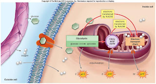
This picture illustrates the production of ATP during cellular respiration- Human Biology, Sylvia S. Mader

Cells can also produce ATP without oxygen through glycolysis and fermentation. These processes are called anaerobic and produce two ATP. Anaerobic systems occur in the cytoplasm and are helpful for short bursts of energy.


I will say that I visited the site with the with the e coli bacteria and ran my mouse over all the reactions. I did find glycolysis but not the kreb's cycle.


E Coli http://biocyc.org/ECOLI/new-image?type=OVERVIEW&force=t

Cells organized into tissue

Tissues are made up of specialized cells, all of the same type, that perform a common function for the body. There are four major types of tissue.

  • Connective tissue

Connective tissue is for binding and supporting body parts. There are lots of kinds of connective tissue. Fibrous connective tissue is found supporting many internal organs like the lungs. Adipose tissue is also fibrous and is used to store fat. Cartilage is a supportive connective tissue and can be found around the nose, and in the outer ear. Bones are the most rigid connective tissue.

I was surprised to learn that blood is a connective tissue, so is lymph.
  • Muscular tissue

Muscular tissue serves to move the body. Muscle fibers make up muscular tissue. There are three types of muscular tissue, Skeletal, smooth, and Cardiac. Cardiac muscle tissue is found only in the heart.

  • Nervous Tissue

Nervous tissue is composed of neurons and neuroglia. Neurons are nerve cells and neurolgia support and nourish the neurons. These cells form the nervous system.

  • Epithelial Tissue

The epithelium is composed of tightly packed cells that form a continuous layer.You will find epithelial tissue lining body cavities and organs.Of particular interest to me was glandular epithelial,. I learned that a gland can be just one epithelial cell or many.This tissue excretes a product which makes it glandular.

Cell junctions

Cell junctions make it possible for tissue to perform a particular function. Cell junctions take place when plasma membranes join in these three ways.

  • Tight junctions

At these junctions adjacent plasma membranes actually join, kind of like a zippper. This causes the layer of cells to become an impermeable barrier You will find these junctions in the tissue lining the kidneys.

  • Adhesion junctions
Cytoskeletal fibers of one cell firmly attach to another.Adhesion junctions are foulnd in tissues subject to mechanical stress.
  • Gap Junctions

When adjacent plasma membranes converge, leaving a tiny gap between them, this is a gap junction. There are gap junctions between cardiac muscle cells.

Organ systems

There are eleven organ systems discussed in the text.

Integumentary system- The skin helps Maintain homeostasis (controls temperature), protects the body, receives sensory imput, and synthesizes vitamin D.

Cardiovascular system-Includes the heart, veins, and arteries. The heart pumps the blood throughout the body.

Lymphatic and immune systems- These systems help defend against disease. They also help absorb fat and control fluid balance.

Digestive system-This system includes the esophagus, intestines, and stomach. These help in digestion.

Respiratory system- Obviously the lungs are part of this system, and help with the intake of oxygen.

Urinary system- Includes kidneys and bladder. THis system excretes wastes.

Skeletal system- The skeletal system supports the body and produces blood cells in the bone marrow.

Muscular system- The muscles move the body, maintain posture, and produce heat.

Nervous system- This system includes the brain, nerves and spinal cord. These organs receive sensory imput, initiate motor output, and help coordinate organ systems.

endocrine system- This system produces hormones, responds to stress, and helps regulate fluid and PH balance.

Reproductive system- The reproductive system produces and transports gametes, It also produces sex hormones.

Conclusion

At the end of his presentation, ProfessorFrolich said that the number of cells in the human body is about one hundred trillion cells (is that more than the national deficit?- I think it is close). Even before learning that number I had already decided that cells blow my mind. Cells are a universe in and of themselves. the more we understand cells and how they work within our bodies, the more equipped we will be to improve our lives.

Sources:

http://biocyc.org/ECOLI/new-image?type=OVERVIEW&force=t

folding.stanford.edu/index.html

Human Biology, Sylvia S. Mader


Sunday, September 14, 2008

Lab Project- Build A Cell, Unit 1


Materials included: Styrofoam molds, spray paint, markers, Christmas tinsel, a plastic ball, construction paper, pipe cleaners. paper plates, sequins, embroidery thread, thumb tacks, tooth picks, and labels.


I decided to create my model using craft supplies. My daughter, a classic "tactile learner," has an endless supply. The actual cell itself was created using Styrofoam models that I spray painted. To illustrate the inner parts, mitochondria, Rough ER, Smooth ER, ribosomes, and Golgi Apparatus, I used a combination of embroidery thread and markers. The flagellum and was created with a pipe cleaner and the lysosome and vesicle were made with thumb tacks. The cell membrane is made out if construction paper. The nucleolus is represented by a plastic ball.


The cell model itself shows the cell in interphase, the stage a cell spends most of its time in. I tried to illustrate a vesicle going through exocytosis, but I do not think I got the full effect. Below you will see a couple of transitional pictures of the cell assembly.


Here I am creating a mitochondria out of embroidery thread... not easy to do.


The cell in progress, no ribosomes yet.


My next step was to illustrate Mitosis. I was inspired by the illustration in the book, pages 382-383. This illustration was instrumental in helping me understand this process. I used paper plates to create a cell and drew a cell membrane. Pipe cleaners with sequins (for centromeres) were the chromosomes. I used pipe cleaners, and markers to show the spindle fibers and asters.
My purpose was to illustrate the different phases of mitosis. The movement of the chromosomes and spindle fibers is shown in the model. At the end, you should be able to see the two duplicate cells.


Illustrating mitosis, a work in progress.


The cell model and mitosis together.


An up close and personal view of my cell model, complete with labels for each part.

Next I worked on DNA replication, transcription to mRNA, and mRNA translation to proteins. For the DNA and mRNA I used pipe cleaners. I illustrated the bases using different colored pipe cleaners. This was time consuming and did not look as nice as I had hoped. I do realize that my double helix is not twisting as it should *sigh*. Do notice that I changed the color of the mRNA base for Uracil (orange), Thymine is blue. The white pipe cleaners are the sugar/phosphate back bones in DNA and I used blue (I ran out of colors) for the mRNA. For translation I used construction paper for the ribosomes and sequins for the polypeptide. The tRNA is green construction paper.

DNA replication, the new strand is on the right.


Transcription to mRNA, mRNA is on the right.

Translation and protein synthesis in the ribosomes.


Cell membrane: construction paper
Cytoplasm: Styrofoam
Nucleus with nuclear membrane: Styrofoam, paint, markers
Nucleolus: Plastic ball
Rough ER: Embroidery thread, marker
Ribosomes: drawn with marker
Smooth ER: Embroidery thread, marker
Golgi Apparatus: Embroidery thread.
Flagellum with microtubules: Pipe cleaner
Lysosomes and Vesicles: Thumb tacks, marker
Mitochondria: Embroidery thread
Chromatin, chromosomes: Pipe cleaners
DNA: Pipe cleaners
mRNA: Pipe cleaners
Enlarged Ribosomes: construction paper
Polypeptide: Sequins

So basically I have illustrated the parts of a cell, cell interphase, mitosis, DNA replication, transcription and translation. The last chapter, 21, was not sinking in with me. Working on this model helped me to get a better "feel" for DNA and RNA structure and function. I had been holding off on the test because I was not getting it. After working on this model I was able to successfully take the test. I guess I am a "tactile learner" as well.


Saturday, September 13, 2008

Online Lab 1- Microscope Simulator


The microscope is an important tool in biology because it allows scientists to magnify cells and small organisms, making it possible to see them close up. Microscopes have made it possible for scientists to discover new organisms, study cell and tissue structure, and fight disease. Microscopes are also important in the medical field. Years ago , while I was working at a public health clinic, we would use a microscope to find out if patients had certain fungal or bacterial infections. Often we would be able to treat patients right away rather than sending specimens to a lab. Obviously, we would not have been able to provide as high a quality of care without a microscope.


I was surprised to learn that the use of magnification and lenses can be traced as far back as ancient Egypt! The credit for the first actual microscope goes to Zacharias Janssen an eyeglass maker from Holland, around 1595. Because of Zacharias' age, some people think his father Hans might actually have invented it (I am choosing to believe it was the whippersnapper myself, I love it when young people do great things). The Janssen's microscopes were simple by today's standards, tubes with lenses at both ends. Depending on the diaphragm openings, magnification ranged from 3x to 9x. English chemist, Robert Hooke later improved on the early compound microscope and is responsible for coining the word "cell." He also wrote Micrographia which would influence inventor Anton Van Leeuwenhoek. Leeuwenhoek's scopes were simple but considered the best in his day. He was also the first person known to describe organisms like bacteria and protozoans.


and is a "tube microscope" with a lens at each end.


The Janssens' microscopes were also important because of the addition of a second lens. the second lens allows for greater magnification by magnifying the image from the first lens. Compound light microscopes today reach magnification levels up to 1000x. The first electron microscope, built in 1931 by Max Knoll and Ernst Ruska, brings magnification even further, 50,000. There are two kinds of electron microscopes, scanning, SEM and transmission, TEM. I had never seen one so below is a picture I found at rsic.puchd.ac.in/em.html


Another kind of microscope is the dissection microscope, used in dissecting specimens.

I got the image above at http://www.hometrainingtools.com/misc/compound%20parts.jpg I thought it was helpful in showing the parts of a microscope.

So, all of this is good to know, but can I actually operate a microscope? Well, I thought so but admittedly, it has been awhile! I very much appreciated the video for the microscope simulator. It is important to prepare the microscope before trying to view a specimen. The condenser knob should be turned all the way up and the rheostat adjusted to ten. the condenser knob is used to adjust the height of the condenser. The iris helps to adjust the angle of the light coming from the top of the condenser. I learned, by using the simulator, that the iris is important in getting a good quality image. The objective is basically the microscope and contains lenses. The nose piece we used in the simulator revolved and had different levels starting at 4x and going all the way up to 100x. The website with the history of the microscope explained magnification as the combination of the eyepiece and the objective. However there was no magnification given for the oculars (eyepiece) on the simulator, so I just used the 4ox objective to get my image. Before beginning, the revolving nose piece should be set at the lowest objective, 4x. It is also important to adjust the oculars so they are both even. Additional adjustments may be necessary before viewing the image. The slide is placed on the stage (the platform that holds the slide) and the stage clip(s) hold the slide in place. The coarse adjuster knob should be adjusted away from you, raising the platform up towards the objective. You are now ready to view the image. Below is the image from the website.


I started by moving the coarse adjuster knob to bring the specimen into view. I then adjusted the fine focus knob and the iris to get a clearer image. The simulator had XY controls that moved the specimen back and forth and and front to back. I then rotated the objectives to get a greater magnification. each time I changed the objective I would adjust the fine focus and iris. I did this of course, while looking through the oculars.

Care and clean up is also important. Slides and lenses should be cleaned with lens paper. Kleenex or paper towels can scratch and should not be used. It is also important to turn the microscope off. A few times the microscope was left on at the clinic and the bulb burned out, not good.

This lab was helpful to me. I would have embarrassed myself if I had tried to use a microscope on my own in class. I noticed I would forget steps when I used the simulator and had to review what I had done. I now have a much better understanding of how to use a microscope.